In the chaos of an acute MI presentation, every second matters. A patient arrives with chest pain and inferior ST elevation. The team knows they need to move fast. And somewhere in that urgency, there is a question that can easily get overlooked: is the right ventricle involved?
Right ventricular infarction complicates roughly half of inferior wall STEMIs to some degree — and in the subset with clinically significant RV involvement, the management approach shifts meaningfully. Aggressive fluid loading. Avoidance of nitrates and diuretics. A different risk profile for hemodynamic instability and in-hospital mortality. Getting the diagnosis right, early, changes what happens next.
The problem is that the standard 12-lead ECG doesn’t include the leads that best capture it.
The Right-Sided Lead Problem
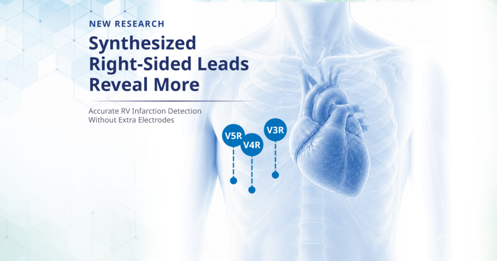
The diagnostic hallmark of RV infarction is ST elevation in the right-sided precordial leads — V3R, V4R, and V5R. These are not part of a standard 12-lead recording. Obtaining them requires repositioning electrodes to the right side of the chest, an additional step that in a fast-moving emergency setting is sometimes delayed or skipped.
There is also a narrow time window to work with. ST elevation in the right-sided leads associated with RV infarction resolves much faster than the ST changes in the limb leads — which means that even a modest delay in obtaining these leads can result in a missed finding. The guidelines are clear: V3R and V4R should be recorded as soon as possible after symptom onset. In practice, the urgency of reperfusion therapy preparation can make that difficult.
What Synthesized ECG Offers
Synthesized ECG technology takes a different approach. Rather than requiring additional electrode placement, it mathematically reconstructs the waveforms of right-sided leads from the data already contained in a standard 12-lead recording — generating a virtual 15-lead ECG without any additional clinical steps.
The concept is not new, but the methods for generating synthesized leads have evolved. Earlier approaches used fixed mathematical coefficients that didn’t account for individual anatomical variation, which could introduce waveform distortions. More recent work has aimed to refine the synthesis algorithms to produce more accurate reproductions of the actual measured leads.
The Study
A recently published study in the Journal of Electrocardiology examined this question directly. Researchers at Nihon University Hospital retrospectively analyzed 38 patients with acute inferior wall myocardial infarction who had undergone both measured and synthesized right-sided precordial ECGs. The study was funded by Fukuda Denshi Co., and used Fukuda Denshi ECG technology to generate the synthesized leads.
The findings were notable. Pearson correlation coefficients between measured and synthesized ECGs were 0.90 for V3R, 0.84 for V4R, and 0.78 for V5R — all statistically significant. Bland-Altman analysis showed negligible bias, with a mean difference of 0.02 mV or less and narrow limits of agreement. For the key diagnostic parameter — ST elevation of 0.1 mV or more in V4R for RV infarction detection — the diagnostic performance of the synthesized and measured leads was statistically equivalent.
The authors concluded that synthesized right-sided ECGs derived from a standard 12-lead recording closely reproduce the measured leads — and may enable rapid identification of RV infarction without adding steps that could delay reperfusion therapy.
Why This Research Direction Matters
The clinical implications of this work extend beyond the specific question of RV infarction. At its core, the study is about what can be extracted from a standard 12-lead ECG that isn’t immediately visible — and whether the limitations of the conventional lead set can be partially addressed through intelligent signal processing rather than additional electrode placement.
That is a question with broad relevance to cardiac diagnostics. Emergency settings where speed is critical. Primary care environments where obtaining extended lead recordings is impractical. Remote monitoring contexts where patient cooperation and technical resources are limited. In each of these settings, the ability to derive more diagnostic information from a standard recording — without additional steps — has real clinical value.
This study is one contribution to that larger body of work. And it reflects something that Fukuda Denshi, as a company, has been investing in for some time: not just building ECG hardware, but understanding what the signal it captures can be made to reveal.
Fukuda Denshi’s Diagnostic Cardiology Portfolio
The research described above was conducted using Fukuda Denshi ECG equipment developed by Fukuda Denshi Co. in Japan. In the United States, Fukuda Denshi USA offers the CardiMax FX-9800 and FX-8400 ECG systems — designed to deliver clinical-grade ECG signal quality in the settings where cardiac evaluation begins. For more information about Fukuda Denshi’s research and technology development, visit fukudaamerica.com/about.
—————
Learn more about Fukuda Denshi’s diagnostic cardiology solutions at fukudaamerica.com.
For more information, contact us at 1-800-365-6668 or [email protected]. You can also reach out to us through our Contact Form.